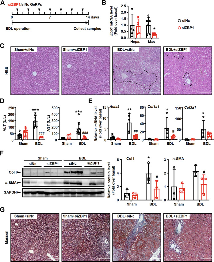
Fig. 9. Specifically knockdown of macrophage Zbp1 alleviated inflammation/fibrosis in BDL liver.
A The schedule of mouse model. B mRNA expressions of Zbp1 in hepatocytes (Hepa.) and macrophages (Mφs) from BDL mouse livers treated with NC or Zbp1 siRNA-GeRPs. C Representative images of H&E staining. Black dashed: inflammation area. Scale bars: 50 μm. D Serum AST and ALT levels were detected in NC and Zbp1 siRNA-GeRPs pretreated BDL livers. E mRNA expressions of fibrosis markers (Acta2, Col1a1 and Col3a1) were detected by qRT-PCR. F Western blot analysis for Col I and α-SMA in the liver tissues from BDL mouse livers treated with NC and Zbp1 siRNA-GeRPs. G Representative images of Masson staining. White dashed: collagen deposition area. Scale bars: 50 μm. Data are presented as the mean ± SEM. *p < 0.05, **p < 0.01, ***p < 0.001 (versus control). #p < 0.05, ##p < 0.01, ###p < 0.001 (versus BDL alone).

