Figure 4.

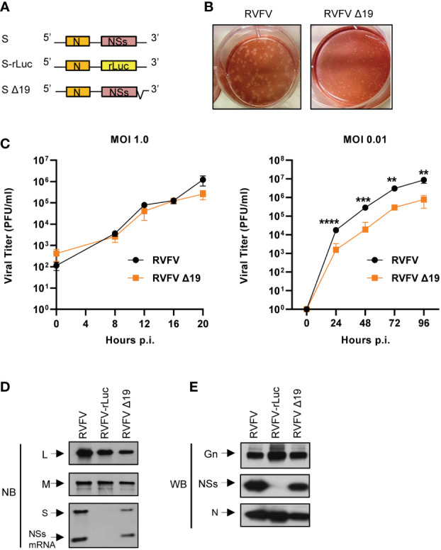
Rescue and characterization of RVFV Δ19. (A) Schematic diagram of S RNA and S RNA mutants in the antigenomic sense. S-rLuc RNA contains the rLuc ORF in place of the NSs ORF. S Δ19 RNA contains a deletion of 19 nt in the 3’-terminal NCR of the antigenomic S RNA. (B) Plaque morphologies of RVFV and RVFV Δ19 in Vero E6 cells. (C) Growth curves of RVFV and RVFV Δ19 in Huh7 cells. Huh7 cells were infected with RVFV or RVFV Δ19 at an MOI of 1.0 (left panel) or an MOI of 0.01 (right panel). Culture supernatants were collected at the indicated time points and the virus titers were determined in Vero E6 cells by plaque assay analysis. The data were obtained from three independent experiments. The values are mean titers and error bars indicate standard deviation. **P < 0.01, ***P < 0.001, ****P < 0.0001. (D) Accumulation of viral RNAs in infected cells. Huh7 cells were infected with RVFV, RVFV-rLuc, or RVFV Δ19 at an MOI of 3. Total intracellular RNAs were extracted at 8 h p.i and equal amounts of intracellular RNAs were analyzed by Northern blot (NB) using digoxigenin-labeled RNA probes that specifically bind to the respective genomic RNA segments. The RNA probe to detect genomic S binds within the NSs ORF resulting in detection of full-length S RNA as well as NSs mRNA. (E) Accumulation of viral proteins in infected cells. Huh7 cells were infected with RVFV, RVFV-rLuc or RVFV Δ19 at an MOI of 3. Cell lysates were collected at 8 h p.i. using the same amount of sample buffer and subjected to Western blot (WB) analysis using anti-Gn antibody (top panel), anti-NSs antibody (middle panel) and anti-N antibody (bottom panel).
