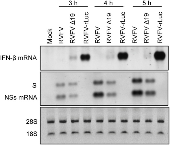
Figure 6.

Accumulation of the genomic S RNA, NSs mRNA and IFN-β mRNA in MRC-5 cells infected with RVFV, RVFV Δ19, or RVFV-rLuc. MRC-5 cells were infected with RVFV, RVFV Δ19, or RVFV-rLuc at an MOI of 3. Total RNAs were collected at various time p.i. as indicated and equal amounts of intracellular RNAs were applied to Northern blot analyses. IFN-β mRNA was detected by using the IFN-β mRNA-specific RNA probe (top panel). Genomic S RNA and NSs mRNA were detected by using a specific RNA probe that binds within the NSs ORF (middle panel). The 28S and 18S ribosomal RNAs (internal loading control) were stained with ethidium bromide, following the separation of the same RNA samples by agarose gel electrophoresis (bottom panel).
