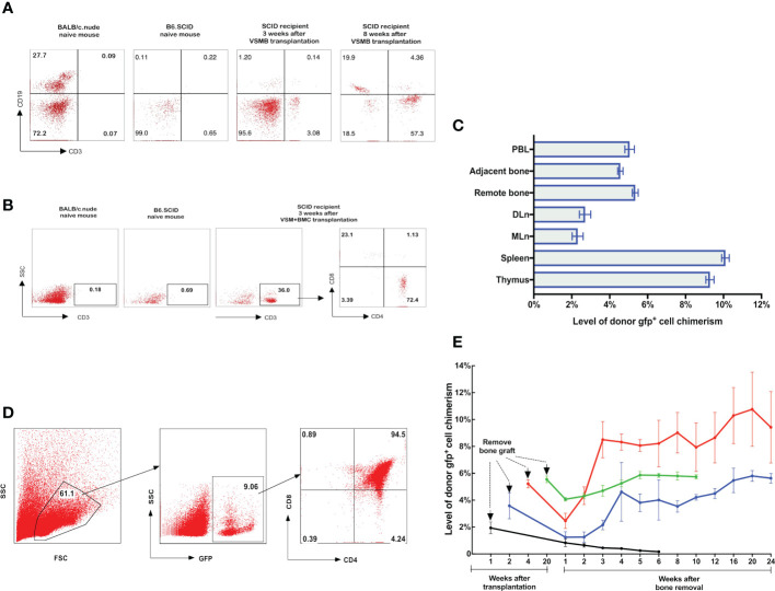
Figure 3.
(A, B) Detection of donor progenitor cells differentiation in SCID mice after receiving VSMB or VSM+BMC from nude mice. VSMB or VSM+BMC (10 x 106) from nude mice were transplanted to SCID mice. CD3+, CD4+, CD8+, and CD19+ lymphocyte sub-populations in blood of SCID mice were assessed by flow cytometry after transplantation. The parent population is lymphocyte, which was gated by forward versus side scatter (FSC vs SSC) plot, followed by analysis of lymphocyte subsets (T and B cells) using CD3 vs CD19 (Fig 3A) or CD3 vs SSC, CD4 vs CD8 (Fig 3B) plots. (A) CD3+ T cells were detected and steadily increased in PBL of SCID recipient at 3 weeks and 8 weeks after VSMB transplantation (n=4). (B) Mature CD3+ T cells were detected in PBL of SCID recipient only at 3 weeks after VSM+BMC. Data are representative of four independent experiments. (C, D) Tracking of donor cells homing in VSMB allograft recipient with sustained stable MC. (C) At 20 weeks post VSMB transplantation from gfp-SD to LEW, the gfp+ cells in PB, DLn, MLn, splenocytes, thymocytes, bone marrow of the tibia adjacent to allograft (adjacent bone) and contralateral tibia (remote bone) of recipients were assessed by flow cytometry. FSC vs SSC plots were used to firstly remove cellular debris and dead cells, and gfp+ subpopulations were then measured for its percentage in the live cells. Data show the results (mean ± SEM) of four recipients. (D) Representative flow cytometric analysis on thymocytes of VSMB allograft recipient at 20-week post-transplantation. Around 9% of thymocytes are donor gfp+ cells, in which gfp+CD4+CD8+ cells account for >90%. (E) The influence of vascularized bone components in inducing and maintaining MC. LEW rats received VSMB allografts from gfp-SD rats and the bone component of VSMB graft was removed from the recipients at 1, 2, 4, and 20 weeks after transplantation. The gfp+ cells in PBL were sequentially assessed by flow cytometry. The level of donor chimerism experienced a transient decrease, then increased to around 6.0-12.0%, and persisted for > 24 weeks after the bone component being removed at 2, 4, 20 weeks, except that at 1 week. Data points represent results (mean ± SEM) for recipients at each time point: week 1 (n=2), week 2 (n=6), week 4 (n=6), week 20 (n=8).

