
Fig. 4.
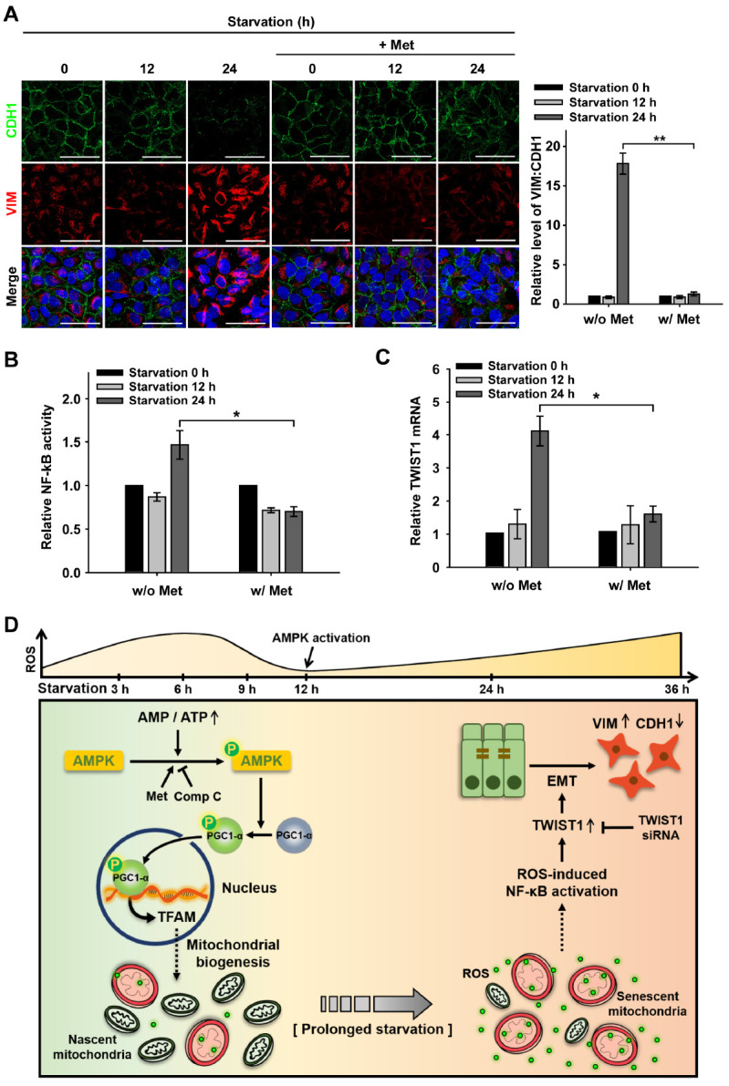
AMPK decelerates EMT by down-regulating TWIST1 under prolonged starvation. ARPE-19 cells were starved with HBSS medium in the presence or absence Met (1 mM; pre-treated for 2 h) for 0-24 h. (A) CDH1 and VIM-immunostained (green and red, respectively) ARPE-19 cells under nutrient starvation for 0-24 h in the presence or absence of Met. Nuclei were stained with DAPI (blue). Scale bar: 50 μm. Bar graph represents the VIM-to-CDH1 fluorescence ratio. Data are presented as the mean ± SEM, n = 3. **P < 0.01. (B) Dual-luciferase reporter assay for NF-κB activation in ARPE-19 cells under nutrient starvation for 0-24 h in the presence or absence of Met. Bar graph represents the activity of NF-κB luciferase which was normalized to Renilla luciferase. Data are presented as the mean ± SEM, n = 3. *P < 0.05. (C) Quantitative real-time PCR for the relative mRNA level of TWIST1 in ARPE-19 cells under nutrient starvation, which was normalized to starvation 0 h in the presence or absence of Met. Data are presented as the mean ± SEM, n = 3. *P < 0.05. (D) Proposed mechanism of the cellular fate of the RPE under nutrient starvation via AMPK activation and the epithelial-mesenchymal transition (EMT).