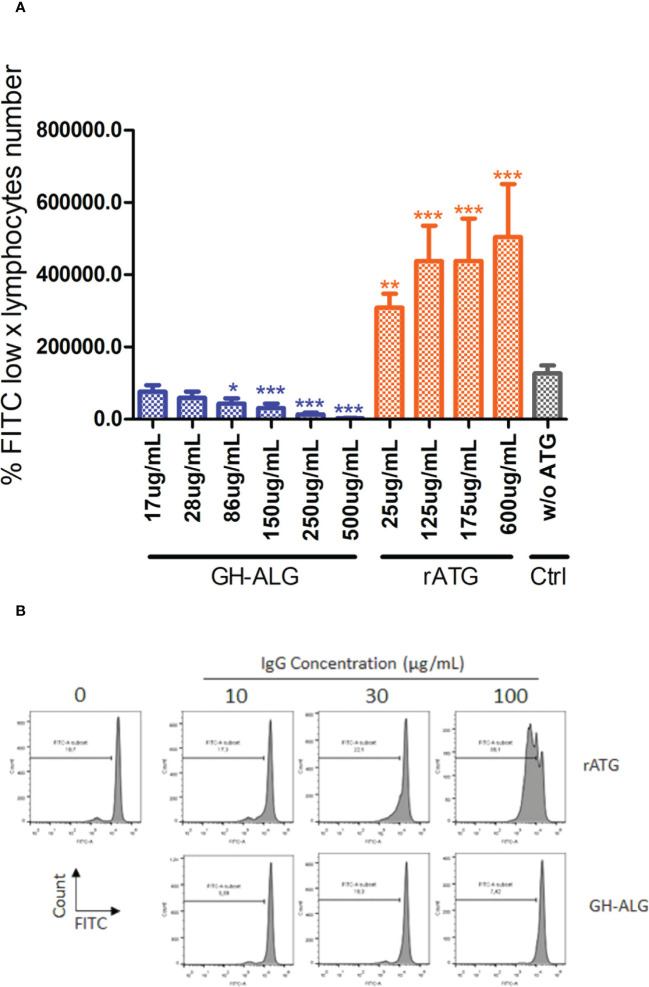
Figure 3.
Mixed Lymphocyte Reaction assay. (A) Stimulator human PBMCs were irradiated with 35 Gy; responder PBMCs were labeled with CFSE. Stimulator cells were adjusted to 8.106 cells/mL, and responder cells were adjusted to 2 × 10⁶ cells/ml of RPMI medium. Next, the PBMCs were cocultured in a total volume of 200 µl of RPMI medium at 37 °C in a 5% CO2 incubator in the dark for 3 days with or without 250 µg/mL of rabbit ATG or GH-ALG. Proliferation was evaluated by measuring CFSE incorporation using flow cytometry. Groups were compared using ANOVA and the Tukey‒Kramer multiple comparison test. (B) Representative flow cytometry histogram demonstrating serial dilution of FITC. * between 0.01 and 0.05. ** between 0.001 and 0.01. *** between 0 and 0.001.

