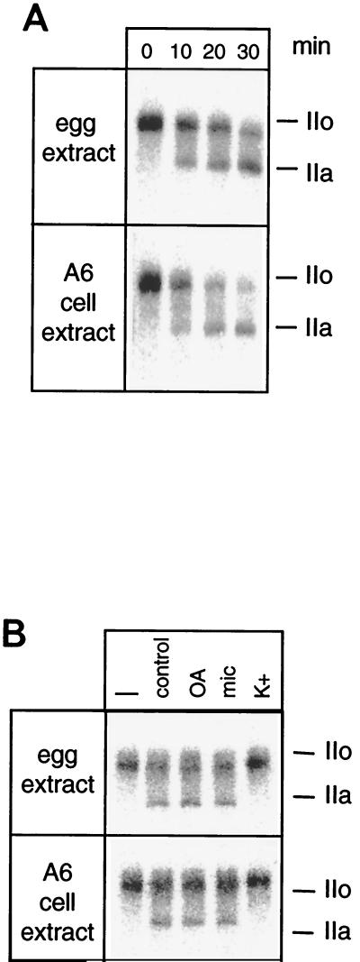
FIG. 1.
CTD phosphatase activity in Xenopus. (A) Interphasic egg extracts or low-salt extracts from Xenopus A6 cells were incubated with labeled RNAP IIO for the amount of time indicated. (B) Extracts were incubated for 15 min with labeled RNAP IIO in the absence (control) or presence of 10 nM okadaic acid (OA), 5 nM microcystin (mic), or 120 mM KCl (K+). The labeled IIO form was incubated in pure buffer (–). Dephosphorylation of the CTD was visualized by SDS-PAGE followed by autoradiography. The positions of subunits IIa and IIo are indicated in both panels.

