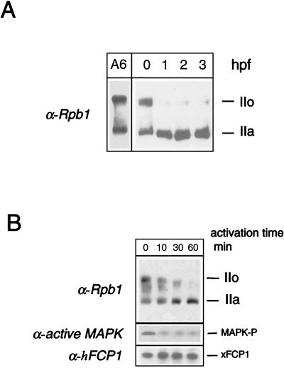
FIG. 5.
CTD phosphorylation state after fertilization in Xenopus. (A) CTD dephosphorylation after fertilization in Xenopus embryos. Batches of 10 unfertilized eggs or embryos were sampled; lysed at 0 (unfertilized eggs), 1, 2, or 3 h postfertilization (hpf); and analyzed by SDS-PAGE along with a whole-cell lysate from A6 cells. The phosphorylation state of the largest RNAP II subunit was analyzed by Western blotting using an anti-Rpb1 antibody. The positions of the subunits IIa and IIo are indicated. (B) CTD dephosphorylation after in vitro activation of a meiotic extract. Activation was performed at 22°C by adding Ca2+ to the CSF-arrested meiotic extract for the indicated time. The phosphorylation state of the CTD and the level of active MAPK were analyzed by Western blotting with anti-Rpb1 and anti-active-MAPK (MAPK-P) antibodies, respectively. The positions of the subunits IIa and IIo are indicated. The presence of xFCP1 was checked by Western blot analysis.

