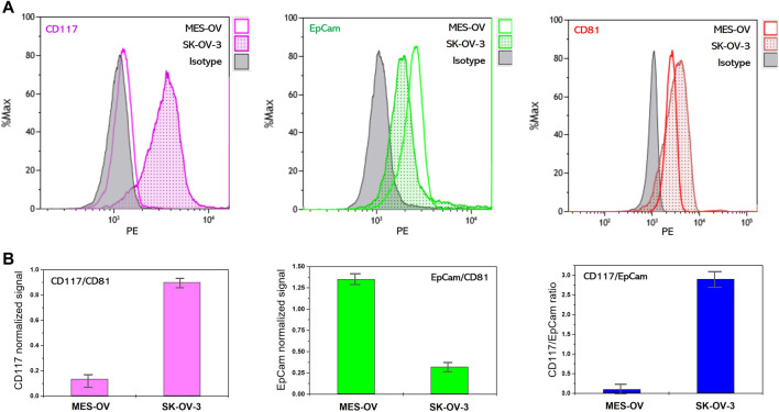
FIGURE 2.
Flow cytometry analysis of membrane protein abundance on MES-OV and SK-OV-3-derived EVs. (A) Flow histograms of CD117-PE, EpCam-PE and CD81-PE staining of MES-OV and SK-OV-3-derived EVs immunoprecipitated on antiCD9 magnetic beads. (B) Ratios of median fluorescent signals. Error bars correspond to the standard deviation of median PE fluorescence calculated from the two measurements.

