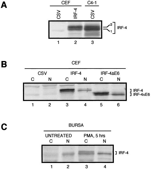
FIG. 3.
Comparison of chicken IRF-4 and IRF-4ΔE6 proteins expressed from retroviral vectors in CEFs with endogenously expressed IRF-4 in chicken B cells. Western blot analysis was performed using IRF-4 antiserum AI4-6. (A) Two isoforms of IRF-4. Lysate from control CSV-infected CEFs is shown in lane 1. Two isoforms of IRF-4 detected in whole-cell lysates of CEF infected with retroviruses expressing IRF-4 (lane 2) comigrate with two isoforms of IRF-4 from whole-cell lysates of the S2A3 v-rel-transformed splenic B-cell line C4-1 infected with CSV (lane 3). Whole-cell extracts from 4 × 10⁵ cells were loaded in each lane. (B) Differential subcellular localization of the IRF-4 isoforms in CEF cultures. Cytoplasmic (C) and nuclear (N) lysates from 2 × 10⁵ CEFs infected with CSV (lanes 1 and 2) or exogenously expressing IRF-4 or IRF-4ΔE6 from retroviral vectors (lanes 3 to 6) were loaded in each lane. (C) Differential subcellular localization of the IRF-4 isoforms in bursal cells. Cytoplasmic (C) and nuclear (N) lysates from normal untreated bursal cells (lanes 1 and 2) and bursal cells treated with PMA for 5 h (lanes 3 and 4). Extracts from 2.5 × 10⁶ bursal lymphocytes were loaded in each lane.

