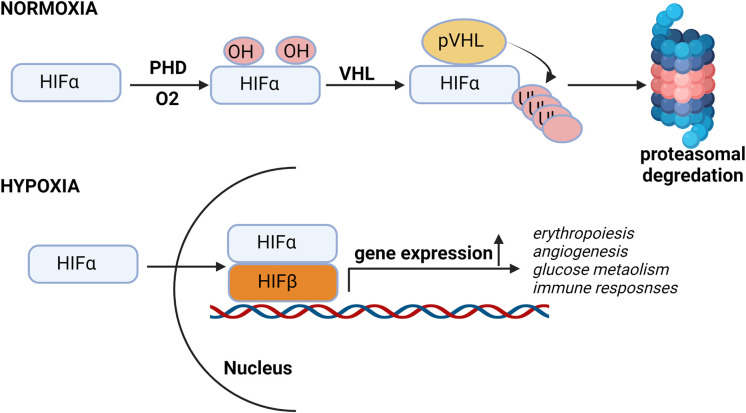
Figure 1.
Hypoxia pathway under normal oxygen and hypoxic conditions. In presence of oxygen, HIF-α subunits are hydroxylated by oxygen-dependent prolyl-4-hydroxylases (PHDs) and then Von Hippel–Lindau protein (pVHL), an E3 ubiquitin ligase, binds to the hydroxylated HIF-α and, which leads to the proteasomal degradation of HIF protein. Under low oxygen conditions, HIF is stabilized and translocated into the nucleus, where it binds to its dimerization partner HIF1β and enhances the transcription of HIF target genes.

