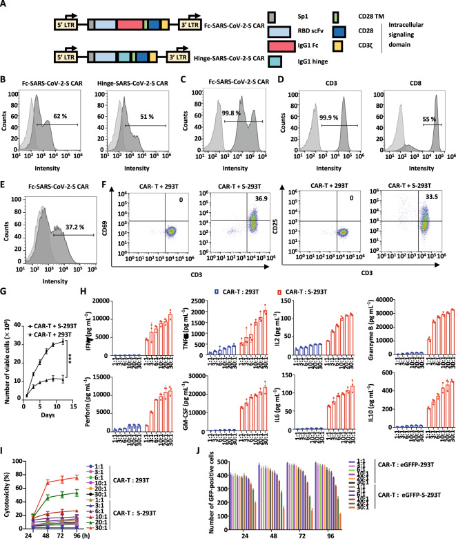
Fig. 1.
The expression, activation, and function of SARS-CoV-2 S protein-targeted CAR-T cells (SARS-CoV-2-S CAR-T). A Schematic illustration of two second-generation SARS-CoV-2-S CAR constructs. The CAR is composed of a signal peptide of the interleukin (IL)2 receptor (Sp1), an anti-RBD scFv, a spacer (IgG1 Fc or IgG1 hinge), a CD28 transmembrane domain (CD28 TM), and intracellular signaling domains (CD28 and CD3ζ). The spacers IgG1 Fc and IgG1 hinge were used in Fc-SARS-CoV-2-S and Hinge-SARS-CoV-2-S CAR constructs, respectively. B 293 T cells transfected with a control lentiviral vector (CTL vector) or SARS-CoV-2-S CAR expression vectors as described in A were stained with eGFP-tagged S protein followed by flow cytometry analysis. Light gray, CTL vector; dark gray, SARS-CoV-2-S CAR. C 293 T cells transfected with a CTL vector or Fc-SARS-CoV-2-S CAR expression vector as described in A were stained with an APC-conjugated anti-human IgG-Fc antibody followed by flow cytometry analysis. Light gray, CTL vector; dark gray, Fc-SARS-CoV-2-S CAR. D Primary T lymphocytes from a healthy donor were expanded and stained with an anti-CD3 antibody conjugated with FITC (CD3-FITC) (left panel) or an anti-CD8 antibody conjugated with APC (CD8-APC) (right panel) followed by flow cytometry analysis. Light gray, blank; dark gray, CD3 or CD8 staining. E Primary T lymphocytes infected with control lentivirus (CTL lentivirus) or Fc-SARS-CoV-2-S CAR lentivirus as described in A were stained with an APC-conjugated anti-human IgG-Fc antibody followed by flow cytometry analysis. Light gray, CTL lentivirus; dark gray, Fc-SARS-CoV-2 CAR-S lentivirus. F SARS-CoV-2-S CAR-T cells were incubated with 293 T cells or 293 T cells transfected with S protein (S-293T cells) at a ratio of 3:1 for two days, and T cells in suspension were separated from adherent 293 T cells and costained with CD3-APC and CD69-PE or CD25-FITC followed by flow cytometry analysis. G SARS-CoV-2-S CAR-T cells were incubated with 293 T or S-293T cells and maintained in culture medium for the indicated durations. The number of viable cells was counted at the indicated time points (mean ± s.e.m, ***P < 0.001). H SARS-CoV-2-S CAR-T cells were incubated with 293 T or S-293T cells at different ratios (1:1, 3:1, 6:1, 10:1, 20:1 or 30:1) for three days before measuring the secretion of cytokines, including IFNγ, TNFα, IL2, granzyme B, perforin, GM-CSF, IL6, and IL10. CTL T cells were used as a negative control (mean ± s.e.m). I SARS-CoV-2-S CAR-T cells were incubated with 293 T or S-293T cells at different ratios for the indicated durations, followed by a cytotoxicity assay (mean ± s.e.m). J SARS-CoV-2-S CAR-T cells were incubated with 293 T cells fused with eGFP (eGFP-293T) or 293 T cells expressing spike fused with eGFP (eGFP-S-293T) at different ratios for the indicated duration, followed by GFP fluorescence detection. The number of GFP-positive cells was counted (mean ± s.e.m)

