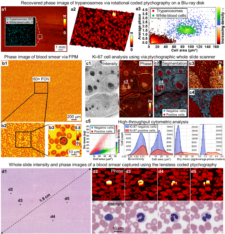
Fig. 7.
High-throughput cytometric analysis via different ptychographic implementations. (a) The recovered whole slide phase image of trypanosomes in a blood smear. The image was acquired using rotational coded ptychography with the specimen mounted on the spinning disk of a Blu-ray drive [61]. (b) The high-resolution recovered phase image of a blood smear using FPM [128]. (c) Ki-67 cell analysis based on the recovered images using the lensless ptychographic whole slide scanner [60]. (d) Whole slide intensity and phase images of a blood smear captured using coded ptychography [30]. The zoomed-in views highlight the phase and intensity images of the white blood cells, which can be used for performing high-throughput differential white blood cell counting.

