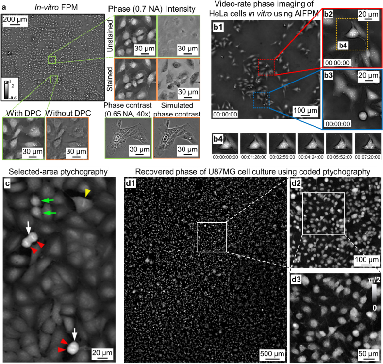
Fig. 9.
2D live-cell imaging via different ptychographic implementations. (a) The recovered phase images of U2OS cell culture using the in-vitro FPM system [122], where the phase is initialized using the differential-phase-contrast approach. (b) Video-rate phase imaging of HeLa cells using annular-illumination FPM [80], where the slow-varying phase information is effectively converted into intensity variations in the captured images. (c) Phase imaging and cell state identification of A549 cells using a lens-based selected-area ptychography system [144]. The white arrows show a proportion of brighter dividing cells, and the intense lines within the cells mark chromosome alignment prior to cytokinesis. (d) The recovered large-scale phase image of U87MG cell culture obtained by a lensless coded ptychography platform [31].

