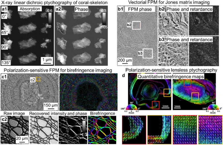
Fig. 11.
Polarization-sensitive imaging using different ptychographic implementations. (a) The recovered absorption and phase images of coral-skeleton particles via X-ray linear dichroic ptychography [296]. (b) The large-area phase and retardance reconstruction of a thin cardiac tissue section using vectorial FPM [250]. (c) The recovered intensity, phase, and birefringence map of a Tilia stem using polarization-sensitive FPM [251]. (d) Recovered birefringence maps of mouse eye and heart tissue using polarization-sensitive lensless ptychography [102].

