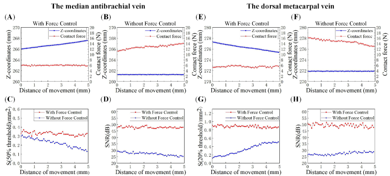
Fig. 8.
Changes of the area and SNR of PA blood vessel images during the force-control and force-uncontrolled scanning of the median antebrachial vein in the wrist (A)-(D) and the dorsal metacarpal vein in hand back (E)-(H). (A) (E) and (B) (F) are the change of the contact force and the spatial coordinate of the probe in the Z direction during the scanning with and without force control, respectively; (C) (G)The cross-sectional areas of the blood vessel region where pixel values larger than the thresholds with the two scanning modes; (D) (H) The SNR calculated with the blood vessel region where pixel values larger than the thresholds.

