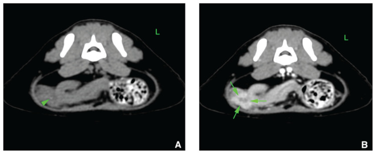
Figure 2.
Computed tomographic view of the caudal abdomen of Case 7 displayed in a soft tissue window in transverse plane (A — No contrast medium injection; B — After contrast medium injection). A 3-mm hyperattenuating linear structure (arrowhead) is seen in the caudoventral, right aspect of the peritoneal space. This structure is located in a slightly hypoattenuating non-enhancing cavitation (arrows), whereas the surrounding soft tissue structures exhibit mild hyperattenuation and enhancement. This structure was interpreted to represent a possible foreign body.

