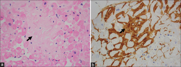
Figure 3.
Histology specimen from endomyocardial biopsy. (a) Photomicrograph shows myocardium with extracellular accumulation of hyalinised material (arrow) in between cardiomyocytes (H&E stain, x200). (b) Photomicrograph shows extracellular material that is immunoreactive for amyloid P protein (arrow) (immunoperoxidase, x200).

