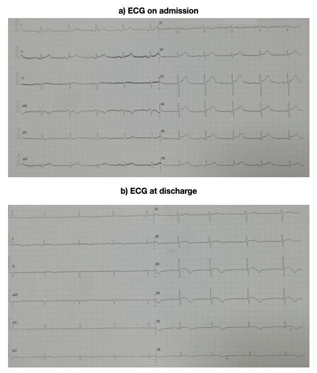
Figure 1 . ECG at admission and at 24 hours. A) ECG on admission: sinus rhythm with a frequency of 61 beats per minute (bpm), the axis was 30°, the PR segment lasted for 0.19 milliseconds (ms), the QRS complex for 100 ms and there was a 0.1 mV elevation of the ST segment in the II and aVF derivations, and a 0.2 mV elevation in the V3, V4, V5 and V6 derivations. B) ECG at discharge: the elevation of the ST segment was still present and the T waves in the V3, V4, V5 and V6 derivations were beginning to become negative.

