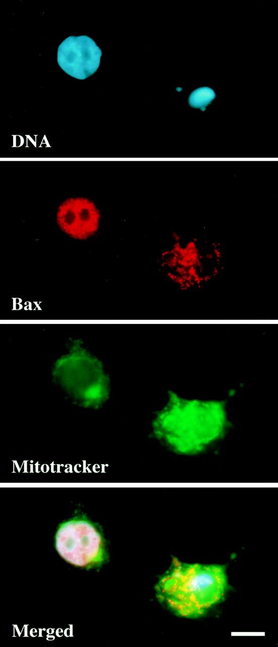
FIG. 3.

LA stimulates the movement of Bax to mitochondria. MCF10A cells were treated with LA (5 μM) for 16 h, fixed in formaldehyde, and immunostained for Bax localization. DNA was stained with Hoescht 33342, and Mitotracker CMX-H2-ROS was added 30 min prior to fixation to label mitochondria. The panels represent three independent images of the same field along with a merged overlay. Bar, 10 μm.
