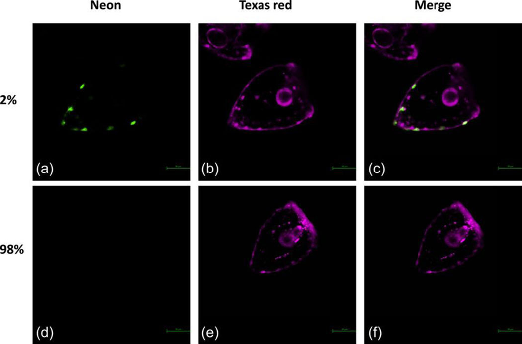
Figure 1: Sp PKS1 Crispr knock-in.
Two percent of embryos express Sp PKS1 Neon in their pigment cells (A). Ninety-eight percent of embryos didn’t show any detectable expression of Sp PKS1 Neon (D). The Texas red dye was co-injected in the zygotes with the knock-in components. This fluorescent dye is used to visualize and sort embryos after injections.

