Figure 3.
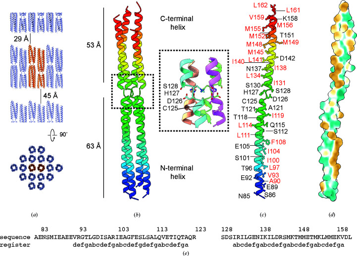
Crystal structure of the BoDV-1 phosphoprotein. (a) Packing of the crystalline lattice showing one asymmetric unit (orange) and neighbouring protein tetramers (blue). (b) Arrangement of the chains in the tetramer coloured from the N-terminus (blue) to the C-terminus (red). The lengths of the two helices and the positions of the helix-breaking motif are annotated. The inset shows the residues involved in the motif, with the four chains coloured uniquely. (c) Residues that stabilize the tetramer by either hydrophobic (red) or electrostatic (black) interactions are annotated. (d) Profile of hydrophobicity across the surface of the protein in the same orientation as in (c). The surface is coloured from hydrophilic (green) to hydrophobic (gold). (e) Repetition of the heptad repeat (a–g) is shown together with the sequence of the oligomerization domain.

