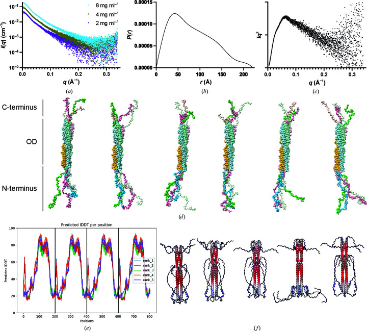
Figure 4.
Flexibility analysis of the BoDV-1 phosphoprotein. (a) Scattering profile, (b) pairwise distribution function and (c) Kratky plot of BoDV-1. The P(r) and Kratky plots are for a protein concentration of 8 mg ml−1. (d) Models showing possible positions of the N- and C-termini generated with CORAL. (e, f) AlphaFold2-predicted IDDT per position and models coloured according to the predicted alignment error from low error (red) to high error (blue).

