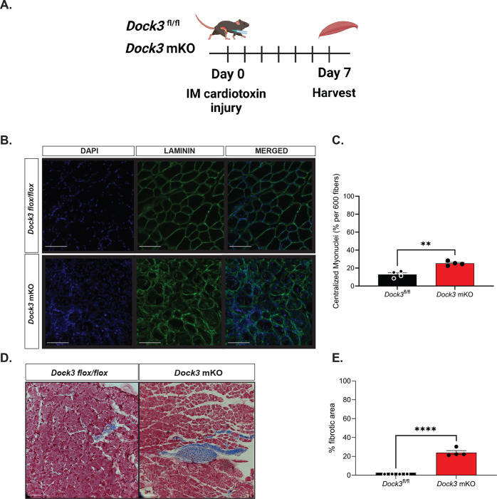
Figure 4. Dock3 mKO mice show impaired skeletal muscle regeneration following injury.
A. Schematic of cardiotoxin induced skeletal muscle TA injury. Dock3 mKO mice and Dock3fl/fl mice were administered with an intramuscular injection of 10 μM of cardiotoxin at Day 0 and sacrificed and harvested day 7 post-injury. B. Cross-section of injured tibialis anterior stained with immunofluorescent antibody against LAMININ (green), DAPI (blue), and the merged image. Scale bar = 200 μm. C. Quantification of immunofluorescent images from (B) analyzing % centralized myonuclei per 600 fibers. **p < 0.001, n = 4 mice/cohort. D. Masson’s trichrome histochemical analysis of injured TA in Dock3fl/fl vs. Dock3 mKO 7 days post-injury. E. Quantification of histochemical images from (D) with analysis of percent (%) fibrotic area in injured TA of Dock3fl/fl vs. Dock3 mKO 7 days post-injury, n = 4 mice/cohort, p < 0.0001. Scale bar = 200 μm.

