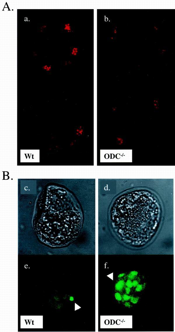
FIG. 5.
ODC is required for cell survival in the ICM. (A) Comparison of the proliferation index between wild-type (a) and ODC−/− (b) E3.5 embryos. Blastocysts were isolated, fixed, and stained with an antibody specific to phosphohistone H3 at Ser10. Two serial sections captured by confocal imaging of each blastocyst are shown. Mitotic cells are readily detected in embryos from both genotypes. (B) TUNEL analysis of E3.5 blastocysts. The upper panels show phase-contrast photographs of wild-type (c) and ODC−/− (d) blastocysts, and the lower panels correspond to immunodetection of TUNEL labeling (e and f). The wild-type embryo displays minimal apoptosis (e). In contrast, the ODC-deficient embryo exhibits massive cell death confined to the ICM (f). The arrowheads indicate fluorescent dots corresponding to fragmented DNA. Wt, wild type.

