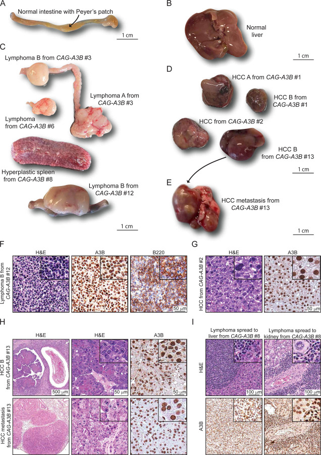
Figure 4. Heterogeneity and evidence for metastasis in tumors from CAG-A3B animals.
(A-B) Representative normal intestine with Peyer’s patch (arrow) and normal liver tissues, respectively, from CAG-A3B mice.
(C-D) Macroscopic pictures of a heterogeneous assortment lymphomas and hepatocellular carcinomas, respectively, from CAG-A3B mice.
(E) Representative image of a primary hepatocellular carcinoma that metastasized to the lung (HCC B from CAG-A3B #13 in panel D).
(F) H&E, anti-A3B, and anti-B220 IHC of lymphoma B from CAG-A3B #12. Inset boxes show the same tumors at 4x additional magnification.
(G) H&E and anti-A3B IHC of HCC from CAG-A3B #2. Inset boxes show the same tumors at 4x additional magnification.
(H) H&E and anti-A3B IHC staining of a primary hepatocellular carcinoma (top) and its metastatic dissemination to the lung (bottom) from CAG-A3B #13. Inset boxes show the same tumors at 4x additional magnification.
(I) H&E and anti-A3B IHC staining of a diffuse large B-cell lymphoma in the liver (left) and kidney (right). Inset boxes show the same tumors at 4x additional magnification.
See also Figures S5 and S6.

