Fig. 4.

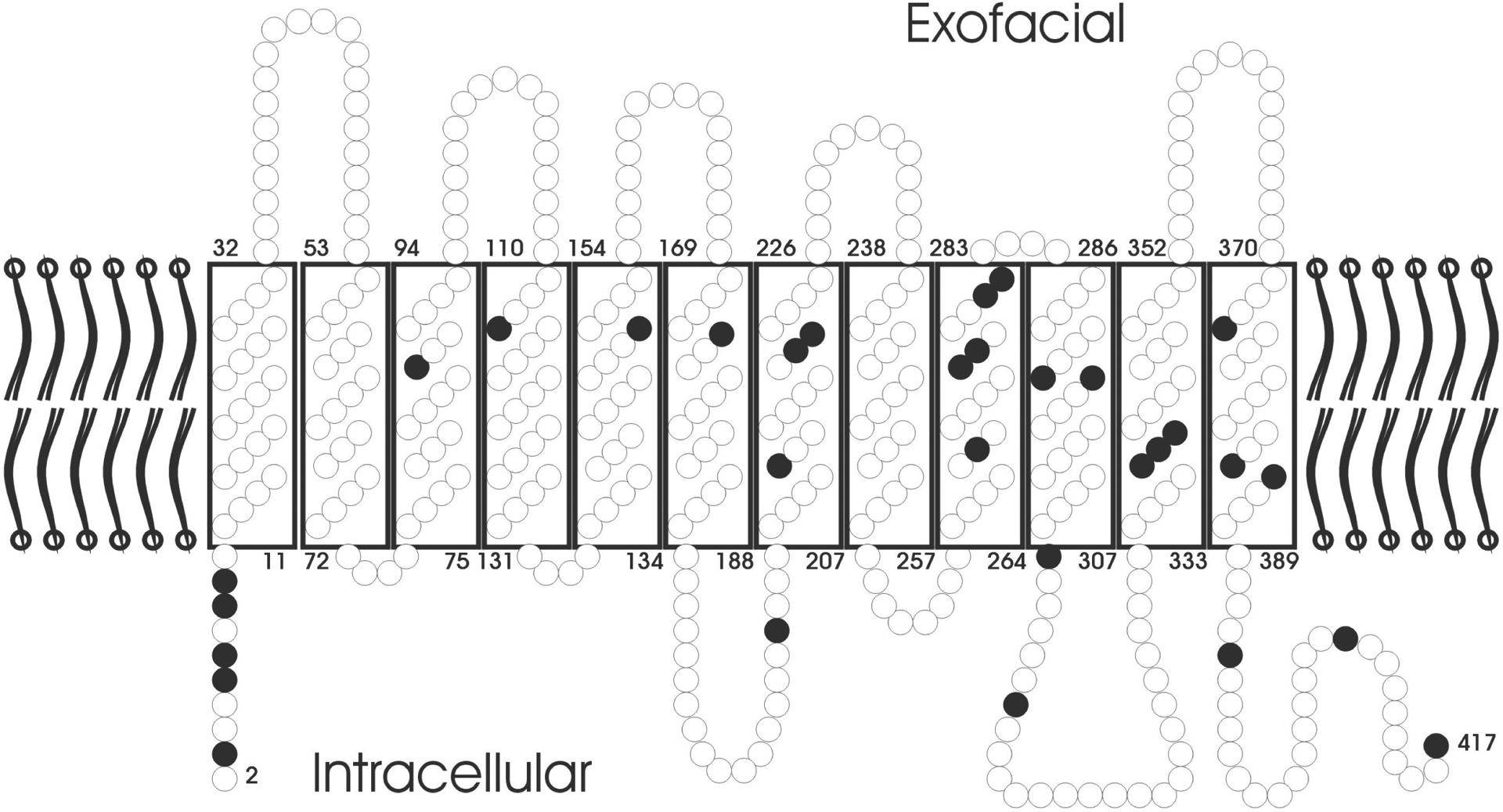
The predicted topology of RhD in the RBC membrane is shown. Amino acids are depicted as circles. Black circles indicate amino acid substitutions, each of which was correlated with a molecularly distinct weak D type.

The predicted topology of RhD in the RBC membrane is shown. Amino acids are depicted as circles. Black circles indicate amino acid substitutions, each of which was correlated with a molecularly distinct weak D type.