
Figure 5.
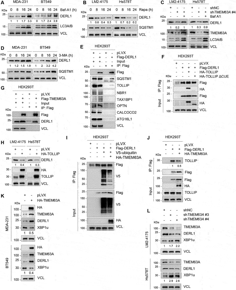
TMEM63A stabilizes DERL1 through blocking TOLLIP-mediated autophagic degradation of DERL1. (A) MDA-231 and BT549 cells were treated with or without 200 nM Baf-A1 for the indicated times and then subjected to immunoblotting analysis with the indicated antibodies. (B) LM2-4175 and Hs578T cells were treated with or without 1 μM rapamycin (Rapa) for the indicated times and then subjected to immunoblotting analysis with the indicated antibodies. (C) LM2-4175 and Hs578T cells stably expressing shNC and shTMEM63A #4 were treated with or without 200 nM Baf-A1 for 24 h and then subjected to immunoblotting analysis with the indicated antibodies. (D) MDA-231 and BT549 cells were treated with or without 1 mM 3-MA for the indicated times and then subjected to immunoblotting analysis. (E-F) HEK293T cells were transfected with the indicated expression vectors, and then subjected to IP and immunoblotting analysis with the indicated antibodies after 48 h of transfection. (G) HEK293T cells stably expressing pLVX and Flag-TMEM63A were subjected to IP and immunoblotting analysis with the indicated antibodies. (H) Immunoblotting analysis of LM2-4175 and Hs578T stably expressing pLVX and HA-TOLLIP with the indicated antibodies. (I-J) HEK293T cells were transfected with the indicated plasmids, and then subjected to IP and immunoblotting analysis with the indicated antibodies after 48 h of transfection. (K) MDA-231 and BT549 cells stably expressing pLVX and HA-TMEM63A were subjected to immunoblotting analysis. (L) LM2-4175 and Hs578T stably expressing shNC and shTMEM63A (#3 and #4) were lysed for immunoblotting analysis. In panels A-D, H, and J-L, densitometric quantitation of western blots was performed using ImageJ.