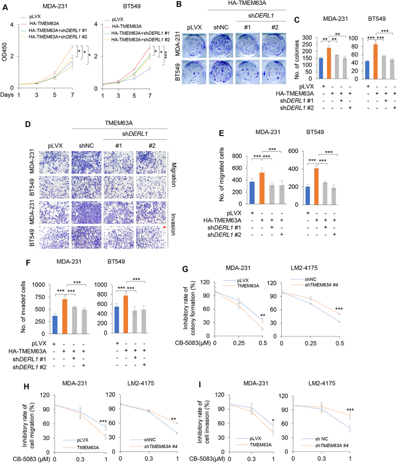
Figure 6.

Pharmacological inhibition of VCP or depletion of DERL1 impairs TMEM63A-mediated TNBC cell proliferation, migration, and invasion in vitro. (A-F) MDA-231 and BT549 cells stably expressing HA-TMEM63A were infected with lentiviral vectors encoding shDERL (#1 and #2), and then subjected to CCK-8 (A), colony formation (B-C), migration (D-E) and invasion (D and F) assays (scale bar:100 μm). Representative images of survival colonies and corresponding quantitative results are shown in B and C, respectively. Representative images of migrated and invaded cells are shown in D, and corresponding quantitative results are shown in E and F, respectively. (G) MDA-231 cells stably expressing pLVX and HA-TMEM63A (left) and LM2-4175 cells stably expressing shNC and shTMEM63A #4 cells (right) were treated with or without increasing doses of VCP inhibitor CB-5083 and then subjected to colony formation assays. Representative images of survival colonies and the relative inhibitory rate of CB-5083 on colony formation ability of MDA-231 and LM2-4175 cells are shown in Supplementary Fig. S5B and Figure 6G, respectively. (H-I) MDA-231 cells stably expressing pLVX and HA-TMEM63A (left) and LM2-4175 cells stably expressing shNC and shTMEM63A #4 cells (right) were treated with or without increasing doses of VCP inhibitor CB-5083 and then subjected to Transwell migration and invasion assays. Representative images of migrated and invaded cells are shown in Supplementary Fig. S5C and S5D. The relative inhibitory rate of CB-5083 on the migration and invasion of MDA-231 and LM2-4175 cells are shown in H and I, respectively. *, **, and *** indicate statistically significant at p < 0.05, p < 0.01, and p < 0.001 level, respectively.
