. 2023 Feb 23;15(1):2171248. doi: 10.1080/19420862.2023.2171248

Figure 4.

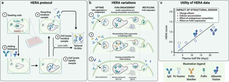
Assay overview. Cells are seeded in plates and, thereafter, biologics are added and incubated during different conditions, followed by analysis of media and cells. These results give a score that correlates with in vivo half-life of the biologic. A) HERA protocol. A plate is shown in the middle with the included steps of the assay illustrated around. 1. A well illustrating an HMEC cell with an endosome inside expressing FcRn is shown. 2. Biologics, depicted as IgG antibodies, Fab fragments, Fc fusions, and a bivalent antibody fragment, are added to the well. 3. The cell is taking up biologics, shown as a cell with an endosome containing biologics. 4. The same cell is depicted, but with the endosome releasing the biologics back to the surrounding media. To the right, a plate well illustrating an ELISA to determine the amount of biologics in the supernatant, or inside the cells, is depicted. B) Three variants of the HERA protocol. To the left, the uptake of biologics through binding to FcRn is shown. Middle, the endosome divides into a lysosome for degradation and recycling of the endosome to the cell surface. To the right, the release of FcRn-bound biologics to the surrounding area is depicted. Three versions of this are illustrated. Top panel shows the use of acidified media resulting in the accumulation and degradation of biologics in the lysosome and no or little recycling of the biologics. Middle panel shows the addition of an antibody binding with its variable region to FcRn, thereby blocking the biologics from binding. This results in the accumulation of biologics in the lysosome and no or little recycling of the biologics. Lower panel depicts various molecules competing for the binding to FcRn, leading to some being degraded and some being recycled. C) A schematic graph illustrating that the score from the HERA assay (y-axes) correlates to the half-life in vivo (x-axes). IgG is in the top right, and IgG with a half-life of above 20 days is shown. In the lower left end, aN Fc-fusion biologic with a half-life of 5 days is depicted. A legend with the various biologics included in the figure, depicted as IgG antibodies, Fab fragments, Fc fusions, and a bivalent antibody fragment, are shown at the bottom.

Human Endothelial Recycling Assay (HERA) as a tool for in vitro pharmacokinetic assessment and addressing FcRn-targeting strategies. A) Generalized HERA protocol. (1) Stably FcRn-transfected human microvascular endothelial cells (HMEC)-1 are seeded, prior to (2) adding FcRn-binding candidate biologics to two parallel cell plates. Following an incubation period, (3) cells from one plate are lysed to obtain an uptake sample. For the other plate, the media is exchanged to recycling medium, and after another incubation period, (4) the medium is harvested as a recycling sample, and (5) the cells are lysed to obtain a residual sample. Candidate biologics in all samples are quantified by an ELISA tailored for specific detection of the assessed biologic. B) Variations of the HERA protocol, enabling analysis of both FcRn-dependent and -independent uptake, cellular accumulation, and FcRn-dependent recycling. Variations include (1) performing the uptake step at mildly acidic extracellular pH, effectively forcing intracellular accumulation of biologics by preventing FcRn-mediated recycling, (2) manipulating FcRn-expression or blocking binding to FcRn to analyze FcRn-dependent and -independent cellular accumulation, and (3) introducing competition for FcRn binding to mirror endogenous competition on the ligand-binding sites of FcRn and its effects on the cellular transport for FcRn-binding biologics. C) HERA data can be used to address the impact of structural design of candidate biologics on FcRn-mediated cellular transport and unspecific cellular accumulation. For some candidate biologics, HERA data may allow for calculation of a score that correlates with plasma half-life in human FcRn-transgenic mice.