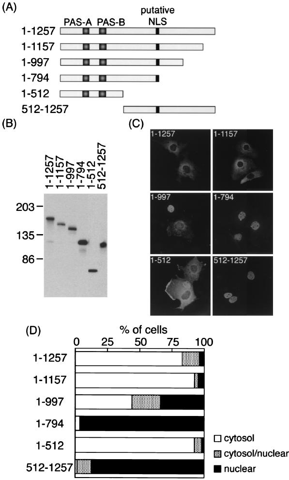
FIG. 1.
Subcellular localization of truncated rPER2 mutants in COS-1 cells. (A) Diagrammatic representation of constructs used to identify the rPER2 NLD. PAS-A, PAS-B, and the putative bipartite NLS are shown as shaded and solid boxes. (B) Confirmation of the size of proteins expressed in COS-1 cells determined by immunoblotting. Sizes are indicated in kilodaltons. Anti-rPER2 antiserum was the first antibody. (C) Representative micrographs show the subcellular localization of rPER2. COS-1 cells were transiently transfected with truncated constructs 1–1257, 1–1157, 1–997, 1–794, 1–512, and 512–1257. Forty-eight hours later, cells were fixed and expressed proteins were visualized using anti-rPER2 antiserum and fluorescein isothiocyanate-conjugated secondary antibody. (D) Quantitative analysis of the above. Subcellular localization was categorized as cytoplasm, cytoplasm and nucleus, and nucleus. The ratio of cells with predominant localization to the total transfected cells was determined by counting 50 to 100 cells three to five times in each experiment under light microscopy.

