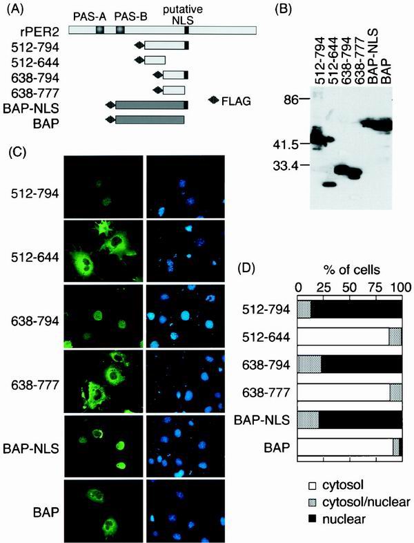
FIG. 2.
Expression of NLDs of rPER2 and rPER2 NLS-tagged BAP (NLS-BAP). (A) Schematic diagrams of four constructs covering the NLD of rPER2. All fragments were tagged with FLAG at the amino terminus. rPER2 NLS was inserted at the carboxyl terminus of BAP. (B) The molecular size in kilodaltons of FLAG-tagged NLD fragments and BAP fragments was confirmed by immunoblotting using anti-FLAG M2 monoclonal antibody. (C) Subcellular localization of rPER2 NLD mutants (512–794, 512–644, 638–794, and 638–777) expressed in COS-1 cells and examined by immunofluorescence microscopy. Fragments of rPER2 NLD mutants were stained with a combination of anti-FLAG monoclonal antibody M2 and fluorescein isothiocyanate-conjugated anti-mouse IgG (green, left panels), and nuclei were visualized with DAPI (blue, right panels). The distribution of NLS-BAP and BAP was also confirmed as described above. (D) Quantitative analysis of above as described in the legend for Fig. 1D.

