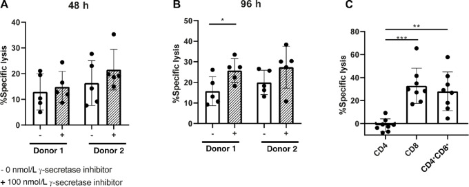
FIGURE 5.
HD T cells kill primary CLL cells in presence of BCMAxCD3 BsAb, which is largely dependent on CD8+ T cells. A and B, Measurement of cytotoxicity after PBMCs of HDs were left unstimulated or stimulated with 100 ng/mL BCMAxCD3 BsAb, in the absence (−) or presence (+) of 100 nmol/L γ-secretase inhibitor. T cells from PBMCs were cocultured were cocultured with primary CLL in a 10:1 E:T ratio for 48 (A) or 96 hours (B) (n = 5). C, Measurement of cytotoxicity of primary CLL cells cocultured with CD4+ or CD8+ or CD4+ and CD8+ (1:1 ratio) in a 5:1 E:T ratio for 96 hours in the presence or absence of 100 ng/mL BCMAxCD3 BsAb (n = 8). The P value was calculated by Wilcoxon test (A), paired t test (B), or repeated measures one-way ANOVA (C). Data are presented as mean ± SD. *, P < 0.05; **, P < 0.01; ***, P < 0.001.

