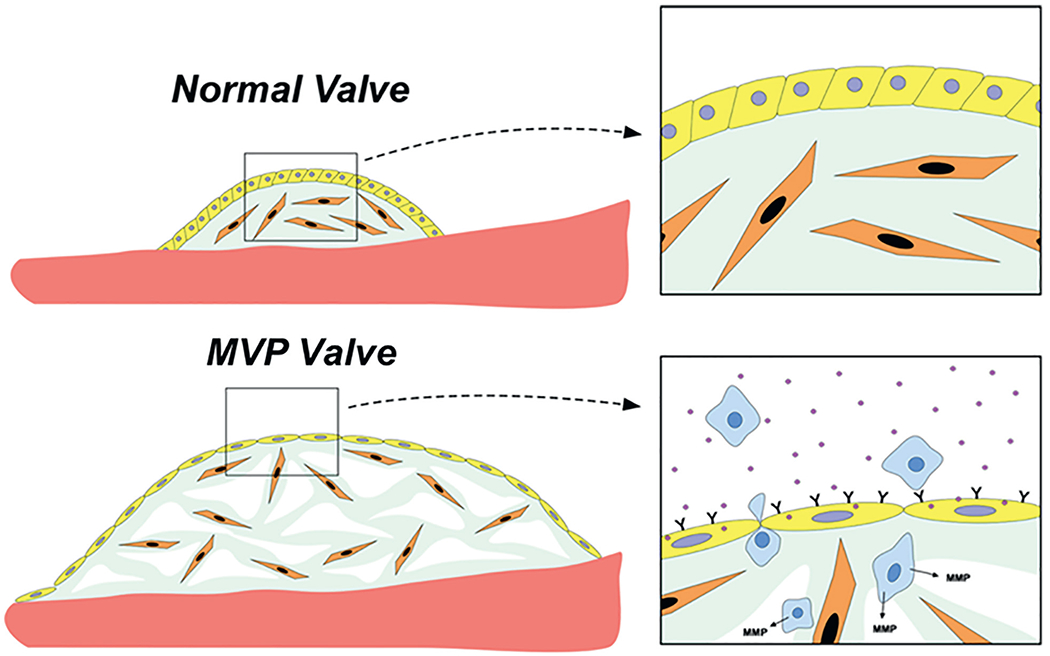
FIGURE 4. Understanding the Developmental Basis of MVP.

Demonstration of the altered interactions between valve endothelial (yellow)/interstitial (orange)/inflammatory cells (blue) using mouse models. MVP = mitral valve prolapse.

Demonstration of the altered interactions between valve endothelial (yellow)/interstitial (orange)/inflammatory cells (blue) using mouse models. MVP = mitral valve prolapse.