FIG. 6.
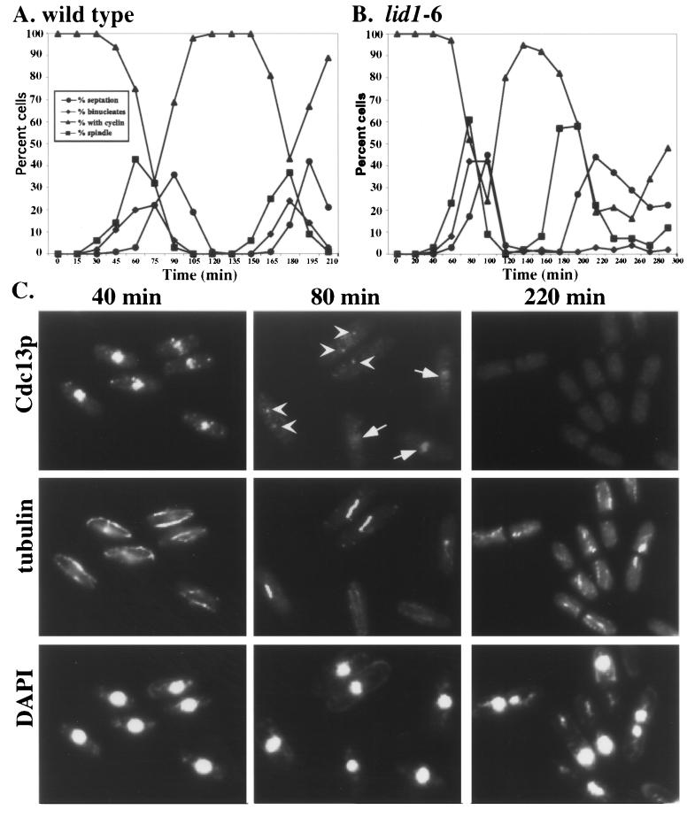
Analysis of Cdc13p localization in APC cut mutants. (A and B) Wild-type (A) and lid1-6 (B) cells were grown to mid-log phase at 25°C in YE medium, synchronized in early G2 phase by centrifugal elutriation, and then shifted to 36°C. Cells were collected at 15-min (for wild-type cells) or 20-min (for lid1-6 cells) intervals and processed for indirect immunofluorescence. The percentage of binucleate cells and septated cells was determined by DAPI staining. The percentage of cells containing spindles was determined by staining cells with the anti-tubulin TAT-1 monoclonal antibody, and the percentage of Cdc13p-positive cells was determined with affinity-purified anti-Cdc13p. (C) Representative fields of lid1-6 cells at the times indicated stained with affinity-purified anti-Cdc13p, TAT-1, or DAPI. Arrows indicate cells containing nuclear Cdc13p, and arrowheads indicate cells with Cdc13p at SPBs.

