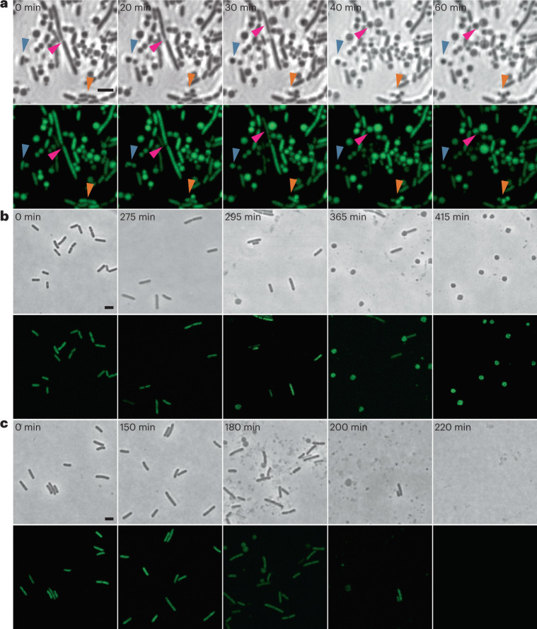
Fig. 1. Phage-induced lysis leads to formation of wall-deficient bacterial cells under osmoprotective conditions.
a, The effects of infection with a lytic phage on the transition of L. monocytogenes Rev2 to wall-deficient bacterial cells under osmoprotective conditions. Cells expressing chromosomally integrated eGFP were challenged with a strictly lytic mutant of temperate Listeria phage A006 lacking the lysogeny control region (ΔLCR) in osmoprotective DM3Φ medium. Turbidity was monitored by spectrophotometry and before lysis, and cells were placed on DM3 agar for time-lapse microscopy (t = 0 min). Shown are phase contrast (PC) micrographs (top) and the corresponding channel for green light emission (bottom). Individual bacterial transition events are denoted by red, orange and blue arrows. Individual frames were extracted from Supplementary Video 1. b,c, Contrary effects of hypotonic vs osmoprotective medium on phage-induced wall-deficient cells. L. monocytogenes Rev2 cells expressing chromosomally integrated eGFP were infected with temperate phage A006 in osmoprotective DM3Φ medium (b) or standard hypotonic 0.5 BHI medium (c). Cells were sampled at several timepoints before and after phage-induced bacterial lysis, followed by microscopic examination on 0.5 BHI or DM3 agar pads, respectively. In hypotonic medium, transition to the wall-deficient state is accompanied by rapid lysis, whereas under osmoprotective conditions, transition to the wall-deficient state is observed and most cells remain intact. Shown are PC images (top) and the corresponding channel for green light emission (bottom). The figures are representative of at least three independent experiments. Scale bars, 2 µm.

