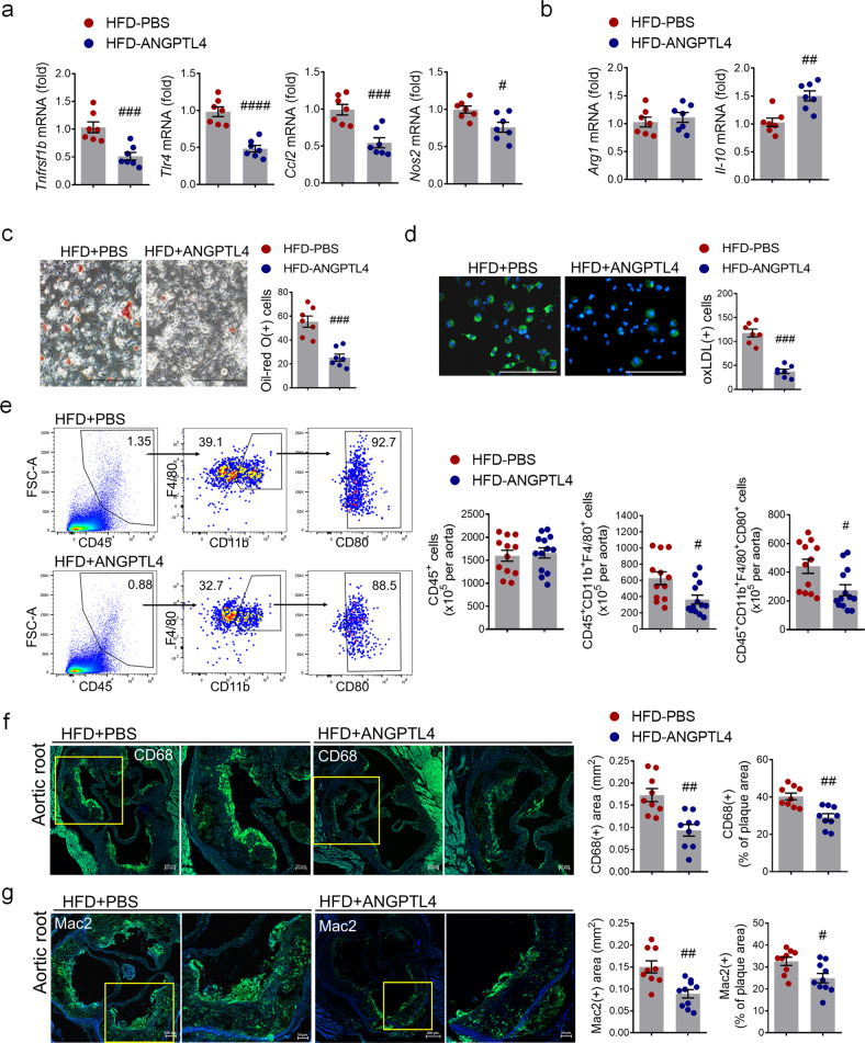
Fig. 2. Effects of ANGPTL4 on macrophage function in atherosclerosis.
a, b Proinflammatory and anti-inflammatory gene expression were analyzed by real-time PCR in BMDMs isolated from Apoe−/− mice treated with PBS or ANGPTL4. c BMDMs from the PBS and ANGPTL4 groups were analyzed using Oil red O staining. Scale bar, 200 μm. d BMDMs from the PBS and ANGPTL4 groups were treated with oxLDL-DyLight 488 for 24 h, and then oxidized LDL uptake was analyzed. Scale bar, 200 μm. e Flow cytometry analyses of single-cell aortic suspensions isolated from the PBS and ANGPTL4 groups. Inflammatory macrophages were quantified by the number of CD80+ cells among the CD45+F4/80+CD11b+ population. f, g The content of macrophages in aortic root sections from the two groups was determined by immunohistochemical staining with anti-CD68 antibody (f) and anti-Mac2 antibodies (g). Representative images are shown, and CD68-positive areas (f) and Mac2-positive areas (g) were measured and quantified as a percentage of the plaque area. Data were presented as the mean ± SEM. #p < 0.05, ##p < 0.01, ###p < 0.001, ####p < 0.0001 (by Student’s t-test).

