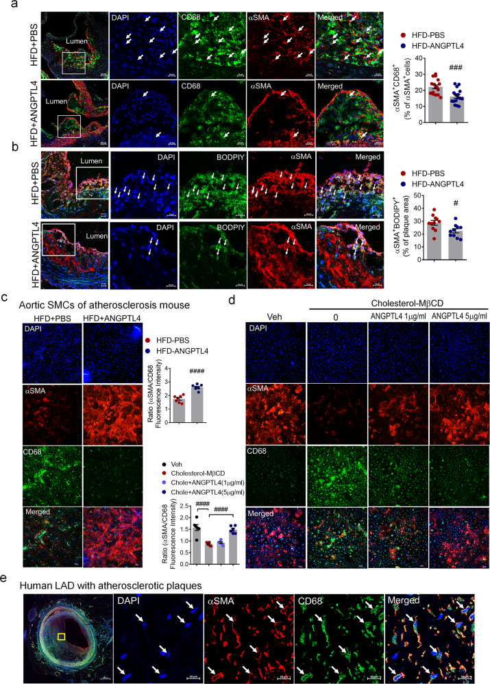
Fig. 5. ANGPTL4 regulates SMC phenotypic changes in atherosclerosis.
a Immunofluorescence staining of atherosclerotic plaques showing CD68 (green) and α-SMA (red) in the aortic root. Boxed areas show close-up images of CD68+α-SMA+ cells (arrowheads) in atherosclerotic plaques. Quantification of the frequency of double-positive (CD68+α-SMA+) cells among the total α-SMA+ cells within the whole lesion and the fibrous cap (n = 16). Scale bar, 20 μm. b Representative images of atherosclerotic plaques in the aortic root showing lipid droplets stained by BODIPY (green) and α-SMA (red). Arrowheads indicate BODIPY+αSMA+ cells. The percentage of BODIPY+α-SMA+ cells within the plaque area (n = 11). Scale bar, 20 μm. c Aortic SMCs were isolated from atherosclerotic Apoe−/− mice treated with PBS or ANGPTL4 and then stained with α-SMA as an SMC marker and CD68 as a macrophage marker. Quantification of α-SMA/CD68 fluorescence intensity. Scale bar, 100 μm. d Aortic SMCs were stimulated with cholesterol (10 μg/ml) for 72 h with or without ANGPTL4 and stained with α-SMA and CD68. Scale bar, 100 μm. e In human LAD, the atherosclerotic lesion displayed cells double positive for α-SMA and CD68. Scale bar, 10 μm. Data were presented as the mean ± SEM. #p < 0.05, ###p < 0.001, ####p < 0.0001 (by Student’s t-test or one-way ANOVA with Bonferroni’s multiple-comparisons test).

