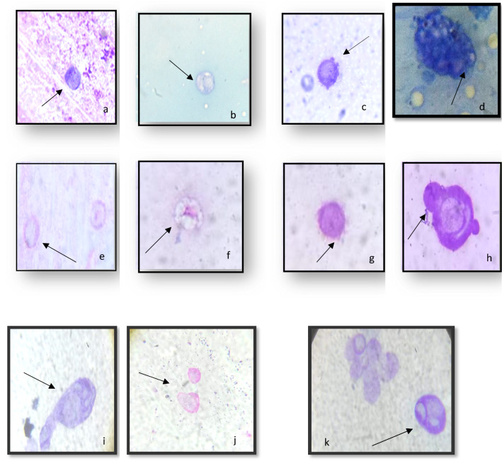
Figure 3.
Effects of exposure to ZnO-NPs at concentrations 50 & 100 μg/ml on hemocyte morphology: (a) Reducing in cytoplasm, (b) Vacuolization in granulocyte, (c) Destroyed cell membrane (d) Numerous vacuoles covering the cytoplasm, (e) Nuclear degeneration, (f) Vacuolization in granulacyte (g) Protruded cytoplasmic contents (h,i), while at concentration100 μg/ml Cytoplasm lysis (j) Apoptotic cells (k) Mitotic division (X100- oil; bar 5 μm).

