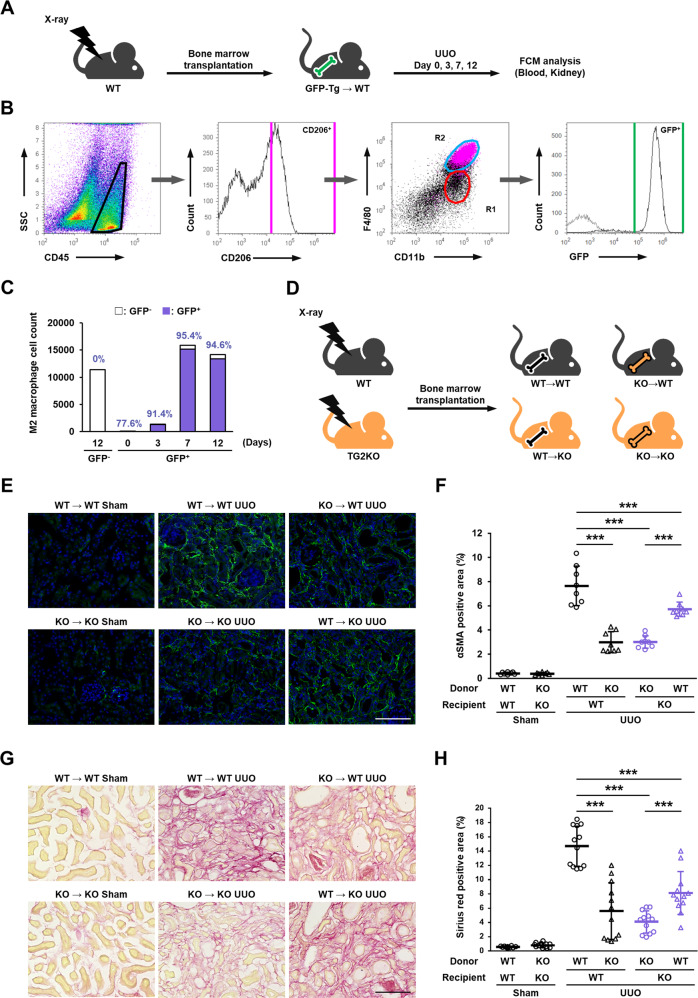
Fig. 3. Role of TG2 in bone marrow-derived cells in renal fibrosis after UUO surgery.
Mice were irradiated at lethal dose (8.5 Gy) of X-rays and transplanted with bone marrow cells isolated from GFP-transgenic mice by tail vein injection. After 4 weeks recovery period, mice were subjected to UUO surgery and analyzed on indicated days (A). The CD45-positive cells in fibrotic kidney were divided into F4/80 and CD11b, and these population classified by CD206 were colored with magenta (B). Then, the counts and percentages of GFP-positive cells in R2 group were analyzed and plotted (C). WT and TG2KO mice were lethally irradiated by X-rays and transplanted with bone marrow cells isolated from WT and TG2KO mice by tail vein injection (D). After 4 weeks recovery period, mice were conducted to UUO and analyzed on 14 days after UUO surgery. The myofibroblasts and collagen fibers in kidney sections were detected by immunofluorescence staining using anti-α-SMA antibody (E) and picrosirius red staining (G), respectively, and the percentages of their positive area are presented (F, H). The nuclei were counterstained with DAPI. Scale bars = 100 μm. Representative results in at least three independent samples were shown. (***P < 0.001 by one-way ANOVA with post hoc Tukey’s multiple comparisons test).

