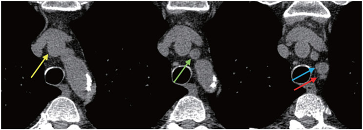
Fig. 2. Axial CT images depicting the identification of the end-aortic arch level.
Left: Only the brachiocephalic artery (yellow arrow) is clearly discernible. Middle: The left common carotid artery (green arrow) is discernible. Right: The brachiocephalic, left common carotid, and left subclavian (blue arrow) arteries become clearly discernible, defining the end-aortic arch level (red arrow).

