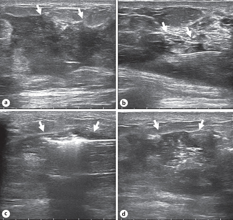
Fig. 3.
Image obtained for a 34-year-old woman who underwent US-guided MWA for NPM in the right breast. Intraoperative parameters: power, 20 W, and ablation time, 299 s. a Ultrasonographic image showing the lesion (arrow). b Water isolation technology (arrow) was used to protect important surrounding anatomical tissues. c Hyperechoic areas in lesions (arrows) seen during ablation. d Ultrasonographic image after ablation shows the lesion area (arrow).

