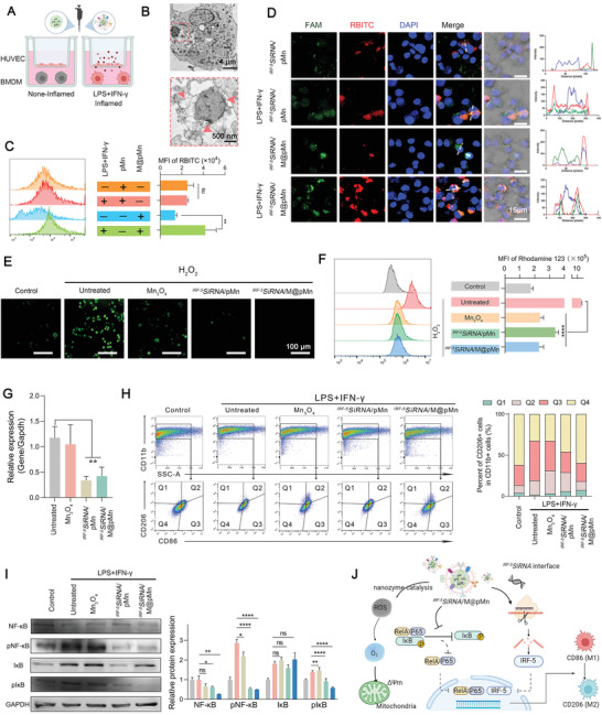
Figure 3.

The biological function of IRF‐5SiRNA/M@pMn on BMDMs includes targeting inflamed macrophages, ROS scavenging, and reducing inflammation. A) Transwell schematic diagram to measure the targeting ability of the nanoparticles. B) Bio‐TEM images of BMDMs cultured with M@pMn (12.5 µg mL−1). C) Fluorescence of pMn and M@pMn under non‐inflammatory and inflammatory BMDMs as measured using FACS (n = 3). D) Representative confocal images of BMDM incubated with FAM‐IRF‐5SiRNA/RBITCpMn and M@ FAM‐IRF‐5SiRNA/RBITCpMn with or without stimulation using LPS and IFN‐γ (Blue, nuclei; red, RBITCpMn; green, FAMsiRNA). E) Fluorescence imaging of BMDMs stained with the DCFH‐DA probe. F) MMP quantification and analysis using flow cytometry (n = 3). G) mRNA expression levels of IRF‐5 as detected via qPCR (n = 5). H) Flow cytometry assay and quantification of BMDM polarization (n = 3). M1 and M2 subtypes are distinguished by the presence of CD86 (mostly in Q3) and CD206 (primarily in Q1). I) Western blotting and quantitative analysis of NF‐κB, pNF‐κB, IκB, and pIκB expression in BMDMs (n = 3). J) Illustration of antioxidant and anti‐inflammatory processes of IRF‐5SiRNA/M@pMn treatment in vitro. Data are presented as mean ± SD. Statistical analysis was performed using a one‐way ANOVA followed by Tukey's post hoc test, *p < 0.05; **p < 0.01; ***p < 0.001; ns, no statistical significance.
