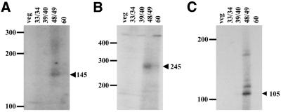
Figure 3.
Spacer-end PCR analyses. Autoradiographs of urea–acrylamide gels containing the extension reaction products from spacer-end PCRs of the left spacer of MacC (A), the right spacer of MacC (B) and the right spacer of RPL-29 (C) are shown. The positions of size standards (in bases) are indicated on the left. The sizes and positions of the expected spacer-end PCR products are indicated by the arrowheads on the right. In each case, the spacer end PCR procedure was performed on total DNA from vegetative cells (lane veg), as well as on total DNA from cells undergoing MAC development at the times (h) indicated (lanes 33/34, 39/40, 48/49 and 60). Images were obtained using a Kaiser RA1 digital camera and AlphaImager 2200 version 5.1 software, and the brightness of images was adjusted using Microsoft PowerPoint software (version 8.0).

