Figure 2.

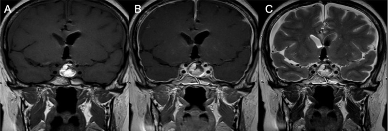
MRI after first surgery: Coronal T1WI (A), Coronal post-contrast T1WI (B) and coronal T2WI revealed signs of intracranial hypotension: right and left subdural collections and pachymeningeal thickening and enhancement.

MRI after first surgery: Coronal T1WI (A), Coronal post-contrast T1WI (B) and coronal T2WI revealed signs of intracranial hypotension: right and left subdural collections and pachymeningeal thickening and enhancement.