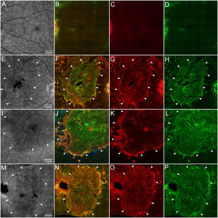
Figure 10.
Retinal glia and vascular changes in areas of RPE atrophy. Aged control (A–D), GA1 (E–H), GA2 (I–L), and GA3 (M–P). Retinas were stained with UEA lectin (white), GFAP (red), and vimentin (green). Aged control retinal flat-mount imaged en face with the ILM uppermost exhibits a uniform vasculature in the posterior pole (A). Control retina imaged en face with the ELM uppermost was nondescript (B–D). GA retinas imaged with the ILM uppermost (E, I, M) exhibit a clear reduction in retinal vascular density in the atrophic area (arrowheads) located in the posterior pole. GA retinas imaged with the ELM uppermost present a large subretinal GFAP/vimentin double-positive membrane-like structure (E–P). Arrowheads indicate the atrophic areas. (A–D) Control, (E–H) GA1, (I–L) GA2, and (M–P) GA3. Scale bars: 1 mm.

