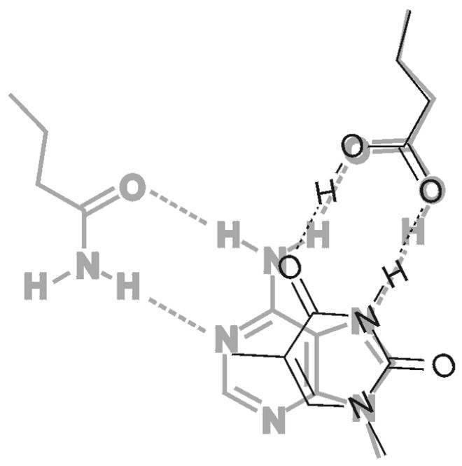
Figure 7.

Model of thymine, bound to acitve site of Mig.MthI (narrow black lines), superimposed onto adenine, bound to active site of MutY.Eco (wide gray lines). The amino acid side chain on the top right is Glu37 in the case of MutY.Eco and Glu42 for Mig.MthI. The amino acid side chain at the top left is Gln182, which corresponds to Leu187 in Mig.MthI (also compare with Fig. 2).
