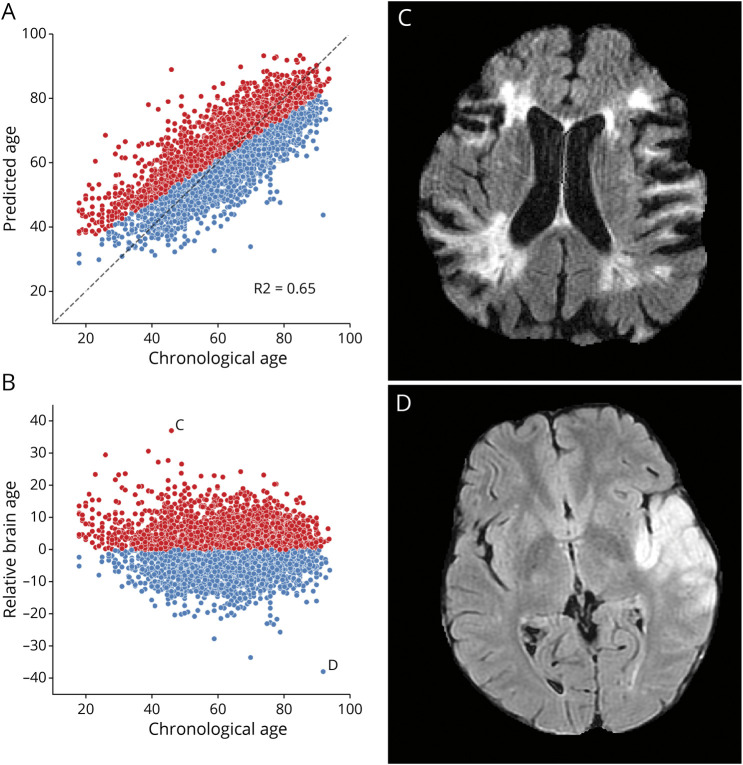
Figure 1. Brain Age Prediction Performances and Relative Brain Age.
Scatter plots of the T2-FLAIR radiomics (A) predicted brain age and (B) relative brain age (RBA) per chronological age. Patients were colored in red if they had a positive RBA and thus a brain that appeared older to their age-matched peers or in blue if they had a negative RBA and a younger-looking brain. (C) T2-FLAIR axial image of a patient with a positive RBA: predicted brain age = 88, chronological age = 46, RBA = 36.2; this patient's brain exhibits multiple cortical and subcortical sequelae, moderate-to-severe parenchymal atrophy with enlarged ventricles and sulci, and confluent white matter hyperintensities, which extents are unexpectedly large for a 46-year-old patient. (D) T2-FLAIR axial image of a patient with a negative RBA: predicted brain age = 43, chronological age = 92, RBA = −38.6; notwithstanding the left middle cerebral artery lesion, this patient's brain trophicity is maintained; the cortex and the deep gray nuclei are sharply defined, overall describing a healthy brain for this 92-year-old patient.

