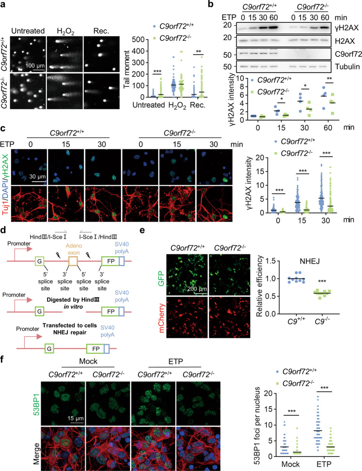
Fig. 2. C9orf72 deficiency leads to attenuated NHEJ repair.
a Impaired DNA damage repair in C9orf72−/− neurons. Primary cultured cortical neurons from C9orf72+/+ and C9orf72−/− mice were treated with 200 μM H2O2 for 3 h (H2O2). After withdrawal of H2O2, the neurons were cultured for 24 h to repair DNA damage (Rec.). DNA damage in neurons was assessed by a comet assay. Quantitative analysis of tail moment. Data are mean (n = 67–237 cells from 3 independent experiments; unpaired t-test; **, p < 0.01; ***, p < 0.001). b Reduced γH2AX level in C9orf72 KO cells compared with WT cells after ETP treatment. HEK293T cells with indicated genotypes were treated with 10 μM ETP for the indicated time points. The γH2AX level was revealed by blotting with anti-γH2AX antibody. Quantitative analysis of γH2AX intensity. Data are mean (n = 4 independent experiments; paired t-test; *, p < 0.05; **, p < 0.01). c Decreased γH2AX level in C9orf72−/− neurons compared with WT neurons after ETP treatment. Primary cultured cortical neurons from C9orf72+/+ and C9orf72−/− mice were treated with 10 μM ETP for the indicated time points, fixed and stained with anti-γH2AX antibody (green) and anti-Tuj1 antibody (red). Quantitative analysis of γH2AX intensity. Data are mean (n = 145–200 cells from 3 independent experiments; unpaired t-test; ***, p < 0.001). d Diagram of NHEJ reporter construct. The NHEJ report construct contains a GFP gene with two HindIII sites in the intron region. The DSB was induced by digestion with HindIII in vitro. e Attenuated NHEJ repair capacity in C9orf72 KO cells. DNA DSB was generated by HindIII digestion in the NHEJ reporter construct. Indicated genotypes HEK293T cells were transfected with cleaved reporter construct. The GFP-positive cells were counted as successful NHEJ repair and mCherry was used for transfection control. Quantitative analysis of relative NHEJ efficiency. Data are mean (n = 10 images of 3 independent experiments; unpaired t-test; ***, p < 0.001). f Reduced number of 53BP1 foci in C9orf72−/− neurons. Primary cultured cortical neurons with indicated genotypes were treated with 10 μM ETP for 30 min, fixed, and stained with anti-53BP1 (green) and anti-Tuj1 (red) antibodies. Quantitative analysis of 53BP1 foci in the nucleus. Data are mean (n = 112–150 cells from 3 independent experiments; unpaired t-test; ***, p < 0.001).

