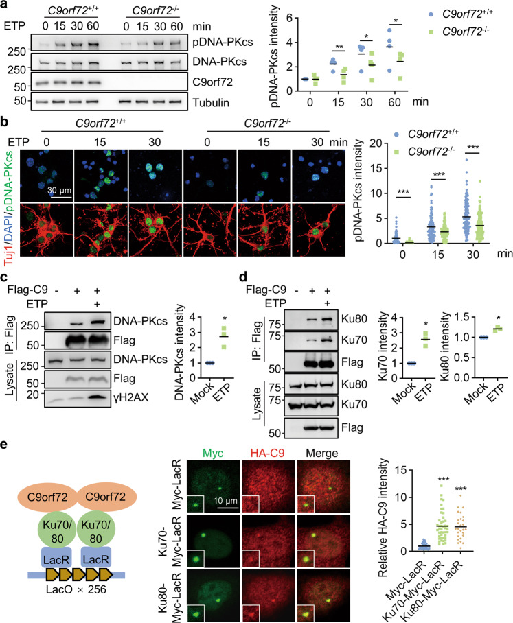
Fig. 3. C9orf72 binds with DNA-PK complex to regulate DNA-PKcs phosphorylation.
a Impaired induction of DNA-PKcs phosphorylation in C9orf72-deficient cells after DNA DSB. HEK293T cells were treated with 10 μM ETP for the indicated time. Cell lysates were blotted for indicated antibodies. Quantitative analysis of pDNA-PKcs intensity. Data are mean (n = 4 independent experiments; paired t-test; *, p < 0.05; **, p < 0.01). b Impaired induction of DNA-PKcs phosphorylation in C9orf72−/− neurons treated with ETP. Primary cultured cortical neurons from C9orf72+/+ and C9orf72−/− mice were treated with 10 μM ETP for the indicated time, fixed, and stained with anti-pDNA-PKcs antibody (green) and anti-Tuj1 antibody (red). Quantitative analysis of pDNA-PKcs intensity. Data are mean (n = 115–150 cells from 3 independent experiments; unpaired t-test; ***, p < 0.001). c, d Increased interaction between C9orf72 and DNA-PK complex after DNA damage. HEK293T cells were transfected with Flag-C9orf72 and treated with ETP. The immunoprecipitated complex was probed with anti-DNA-PKcs antibody (c), and anti-Ku70, and anti-K80 antibodies (d). Quantitative analysis of intensities. Data are mean (n = 3 independent experiments; paired t-test; *, p < 0.05). e Schematic diagram of the LacO/LacR system to analyze C9orf72 interaction with Ku70 and Ku80 on chromatin (left). U2OS LacO cells were transfected with HA-C9orf72 and Ku70-Myc-LacR or Ku80-Myc-LacR, and stained with anti-Myc (green), and anti-HA (red) antibodies. Quantitative analysis of HA-C9orf72 intensity. Data are mean (n = 27–44 cells from 3 independent experiments; one-way ANOVA; ***, p < 0.001).

